

Some technologies that can save lives are already here, and we simply don’t use them. Not even to help unborn babies come into the world without suffering, without complications that may stay with them for the rest of their lives. To save them.
These days, fortunately, we talk a lot more about providing incubators and equipment to save the littlest ones in their hardest moments. But are we doing everything we can to prevent such situations, to make them rarer?
Not according to our guest today—and he knows what he’s talking about.
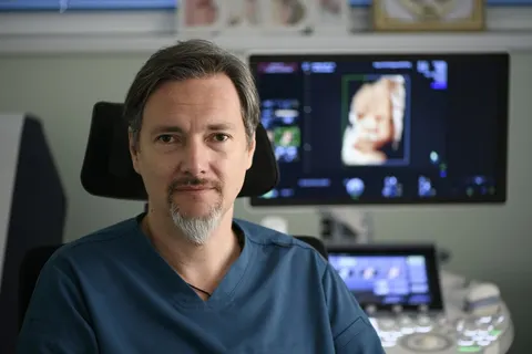
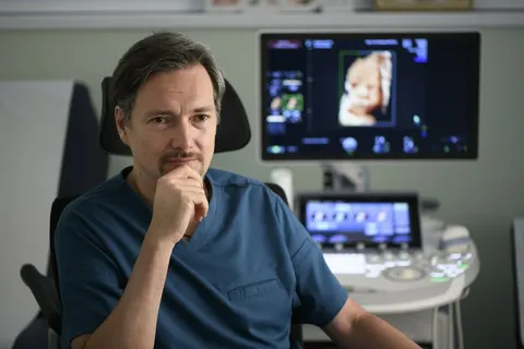
“There are so many ways to help unborn babies, I wish they were talked about much more!” says Assoc. Prof. Petar Ignatov, an obstetrician-gynecologist. Besides his wealth of everyday experience, he is the creator of some impressive innovations. The latest of them could drastically improve how successfully doctors use one of the most important diagnostic methods. What’s more, it has the potential to help diagnose and save a huge number of unborn kids.
Can there be anything more important than taking care of the children to come?
The Child in You
Not long ago, all eyes were fixed on the fate of five people who ended up in the depths, helpless, in the uncertain embrace of the future. That was the Titan submersible, but every one of us has been in a very similar role, even if we don’t remember it.
The last trimester of pregnancy is the time when doctors still speak of a “fetus,” although what is inside is in fact an almost “finished” baby. The big risks of pregnancy and miscarriage are supposedly behind us. Yet it is precisely in this period that many of the problems leading to serious complications occur. And catching them in time can be a matter of life and death.
“Lately the emphasis has been on buying incubators and equipment to help resuscitate newborns and treat complications. But in my view we should also talk about how important prevention is, and how in practice it doesn’t happen,” says Dr. Ignatov.

Child’s Play
“It is very important to add more diagnostic options. So that the condition of babies is assessed before birth, and there are far fewer newborns with complications, with oxygen deprivation causing cerebral palsy and neurological problems,” the doctor adds.
Today Bulgaria’s National Health Insurance Fund covers a small number of mandatory checkups and tests during pregnancy. Meanwhile, obstetrician-gynecologists themselves recommend fetal morphology as an additional exam—a mandatory check in almost all developed countries. This safe, noninvasive method detects a large share of the anomalies and problems that pose a risk to the baby-to-be.
And while most future parents do get this exam (those who can afford it and have found a responsible gynecologist to follow the pregnancy), there are many other important checks.
The pregnancy has reached week 28, the last, decisive third. The tests for chromosomal anomalies and the fetal morphology have been done, and they have shown that the baby is developing normally. The most important question now is how it is feeling in its temporary home. Alas, like the passengers of a submersible, it has no way of telling her, and it is important for doctors to follow the signals. Which, however, rarely happens.

The Unborn Hero
“In the last trimester, the most important thing is to monitor the condition of the fetus, because a number of factors, inside or outside the womb, could have a negative effect. They could lead to fetal distress, as we call it. A lack of oxygen to the brain and vital organs, anemic syndrome, reduced oxygen saturation of the blood,” says Assoc. Prof. Ignatov.
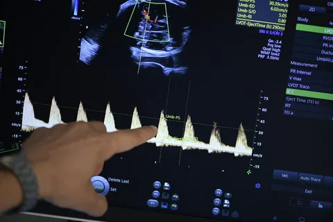
Doppler ultrasound is used in many fields where it is important to track the movement of blood through a vessel. In the last stage of pregnancy it is a fitting solution, harmless and noninvasive, for checking the parameters of blood flow in the baby’s middle cerebral artery.
The method is simple and proven to show whether everything is fine… and yet it is hardly ever used in Bulgaria, which is why complications so often arise.
“Nobody talks about it, and few colleagues are able to apply the method. I very much hope patients will inform themselves, take an interest for the sake of their babies, and as a result more colleagues will want to learn,” the specialist says. He is ready to train his colleagues entirely free of charge, as long as they are willing. This type of examination is already part of the courses for specialists held at the medical center where he works.
Children of Mother Earth
“I want to reach these people, for them to understand that it is not up to God whether the baby survives the third trimester—there are ways to find out what its condition is. If it turns out there are signs of fetal distress, of oxygen deprivation, there are many things that could be done! As a last resort, it can be delivered earlier, but that could save its life!” the obstetrician-gynecologist adds.
Either way, for now the examination is performed in a very small percentage of the roughly 60,000 pregnancies a year in Bulgaria.
And although this convenient method is already quite revealing… here we come to the contribution of Assoc. Prof. Ignatov and his team toward making it, and every other form of Doppler examination, considerably more precise.

Math for Doctors
“For the 40 years that Doppler examinations have existed, they have been interpreted with one and the same formula. Our team proved that it is wrong and that it is time to replace it with another, much more accurate one,” the doctor explains.
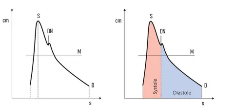
When blood flow through a vessel is monitored, the line reaches an obvious peak before heading down. For a long time, that was believed to be where the line between systole and diastole runs—systole being the moment the heart pushes the blood out and it passes through the vessel under rising pressure, diastole the moment the heart relaxes and the pressure in the vessel falls. The pressure at this peak, referred to as the “pulsatility index,” is the indicator doctors in various fields track. Including in examinations during the last third of pregnancy.
Sometimes, though, what matters most is invisible not just to the eye, but to the instruments as well. In recent years it has become clear that the boundary between systole and diastole is not the obvious peak! Shortly after it, once the pressure has started to drop, there is always a second, lower peak, and that is the true boundary.
Eyes Wide Shut
Although this phenomenon has been proven, medicine is slow to get used to the change. And now Assoc. Prof. Ignatov and his team are offering another important solution, one with the potential to make diagnosis far more accurate!
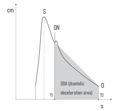
Over the years he has examined many pregnant women, and at first he noticed a correlation intuitively: the larger the area under the curve after the second peak, the greater the risk of a problem.
A month ago, after years of research, his scientific paper on the subject came out. In it, Assoc. Prof. Ignatov and his team propose a formula for calculating the area of the shape formed after the second peak—in diastole.
The pattern: in unborn babies whose shape falls within the normal range, the pregnancy ends calmly. Outside it are children in whom further tests confirm a problem, and timely, adequate measures are needed.
Interest in the topic is high; the paper has already been cited and discussed by professional communities around the world. And Assoc. Prof. Ignatov is preparing the next big step—an international study across different countries and hospitals, involving tens of thousands of patients who would be followed up, so that the new formula can be proven conclusively and turned into the gold standard.
The Bottom Line
But how could no one, for decades, have noticed that the most accurate indicator was not being used to analyze Doppler examinations?
“This boundary really has been misread for the last 40 years,” the doctor says. “It may sound strange, but in medicine it’s not unprecedented. There are many cases where we did something wrong for a long time, or used an unsuitable method. The phenomenon was right in front of our eyes for decades, and no one thought to use it. I, too, didn’t ask myself critically for a long time whether all this held up. Until at some point, like most ideas that come to people, I slapped my forehead. ‘Well, what if…?’ What followed was 8 years of work. We had to handle it purely mathematically and methodically, to recruit patients. And to prepare the publication so that it met the extremely high standards of today’s renowned medical journals in our field.”
Pressure Cooker
“This area measurement is a qualitatively new approach to interpreting all Doppler examinations,” the doctor says. “Introducing this indicator is a huge moment for us, because it changes the whole paradigm in the fields of medicine that deal with assessing blood flow. That’s 70 percent of medical specialties, and veterinary ones too.
This phenomenon, called the ‘diastolic deceleration area,’ exists in all vessels relatively close to the heart—in the head, the arteries to the arm, the pulmonary and renal arteries, even the uterine arteries. So all specialties, such as cardiology, gastroenterology, endocrinology, internal medicine and pulmonology, stand to benefit from studying how this indicator could be useful in their practice,” the doctor sums up.
The method has been patented here, and Assoc. Prof. Ignatov is determined to do everything possible for it to remain a Bulgarian patent.
This is not his first life-saving innovation. For almost a decade now, doctors have been using his platform for automated assessment of the results of another important examination during pregnancy—cardiotocography.
As Assoc. Prof. Ignatov himself says, what matters from here on is finding more like-minded people—among expectant mothers, and as a result among his colleagues. Because many, many lives of unborn babies could be saved.
Can there be anything more important than that?